Ficheiro:Nematocyst discharge.png
Sem resolução maior disponível.
Nematocyst_discharge.png (480 × 371 píxeis, tamanho: 190 kB, tipo MIME: image/png)
|
|
Esta imagem provém do Wikimedia Commons, um acervo de conteúdo livre da Wikimedia Foundation que pode ser utilizado por outros projetos.
|

|
Esta imagem de biologia (ou todas as imagens neste artigo ou categoria) deveriam ser recriadas usando gráficos vectoriais, como ficheiros SVG. Isto tem várias vantagens; veja as Commons:Media for cleanup|imagens para rever para mais informações. Se já criou um ficheiro SVG desta imagem, por favor, carregue-o. Depois do novo ficheiro SVG ter sido carregado, substitua aqui esta predefinição pela predefinição {{vector version available|nome da nova imagem.svg}}.
|
Descrição do ficheiro
| Descrição |
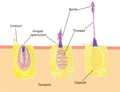
English: The diagram above shows the anatomy of a nematocyst cell and its “firing” sequence, from left to right. On the far left is a nematocyst inside its cellular capsule. The cell’s thread is coiled under pressure and wrapped around a stinging barb. When potential prey makes contact with the tentacles of a polyp, the nematocyst cell is stimulated. This causes a flap of tissue covering the nematocyst—the operculum—to fly open. The middle image shows the open operculum, the rapidly uncoiling thread and the emerging barb. On the far right is the fully extended cell. The barbs at the end of the nematocyst are designed to stick into the polyp’s victim and inject a poisonous liquid. When subdued, the polyp’s tentacles move the prey toward its mouth and the nematocysts recoil back into their capsules. |
| Data | 11 de abril de 2007 (data de carregamento original) |
| Origem | Transferido de en.wikipedia para a wiki Commons. |
| Autor | Este ficheiro foi inicialmente carregado por Spaully em Wikipédia em inglês |
Licenciamento
Este ficheiro está sobre a licença Creative Commons CompartilhaIgual 1.0.
A Creative Commons reformou este instrumento legal e não recomenda que seja aplicado a trabalhos.
|
Esta imagem está em domínio público pois ela contém material que vieram originalmente da National Oceanic and Atmospheric Administration dos EUA, tirada ou feita durante o trajeto de um funcionário em obrigações oficiais.
|
Registo de carregamento original
A página de descrição original está aqui. Todos os nomes de utilizador a seguir referem-se a en.wikipedia.
- 2007-04-11 17:10 Spaully 480×371×8 (194868 bytes) Modified from: http://www.oceanservice.noaa.gov/education/kits/corals/media/supp_coral01b.html {{Information |Description=Nematocyst discharge process. |Source= Modified from [http://www.oceanservice.noaa.gov/education/kits/corals/media/supp_coral01b.html
Histórico do ficheiro
Clique uma data e hora para ver o ficheiro tal como ele se encontrava nessa altura.
| Data e hora | Miniatura | Dimensões | Utilizador | Comentário | |
|---|---|---|---|---|---|
| atual | 17h29min de 13 de outubro de 2007 | | 480 × 371 (190 kB) | Alison | {{Information |Description===Description== The diagram above shows the anatomy of a nematocyst cell and its “firing” sequence, from left to right. On the far left is a nematocyst inside its cellular capsule. The cell’s thread is coiled under pressur |
Utilização local do ficheiro
As seguintes 2 páginas usam este ficheiro:
Utilização global do ficheiro
As seguintes wikis usam este ficheiro:
- ca.wikipedia.org
- ceb.wikipedia.org
- en.wikipedia.org
- fr.wikipedia.org
- hr.wikipedia.org
- id.wikipedia.org
- it.wikibooks.org
- ja.wikipedia.org
- lv.wikipedia.org
- ms.wikipedia.org
- my.wikipedia.org
- pa.wikipedia.org
- simple.wikipedia.org
- sv.wikipedia.org
- te.wikipedia.org
- th.wikipedia.org
- vi.wikipedia.org