Ficheiro:Anatomy of Human Ear with Cochlear Frequency Mapping.svg

Dimensões desta antevisão em PNG do ficheiro SVG: 674 × 519 píxeis Outras resoluções: 312 × 240 píxeis | 624 × 480 píxeis | 998 × 768 píxeis | 1 280 × 986 píxeis | 2 560 × 1 971 píxeis.
Imagem numa resolução maior (ficheiro SVG, de 674 × 519 píxeis, tamanho: 33 kB)
|
|
Esta imagem provém do Wikimedia Commons, um acervo de conteúdo livre da Wikimedia Foundation que pode ser utilizado por outros projetos.
|
Descrição do ficheiro
| Descrição |
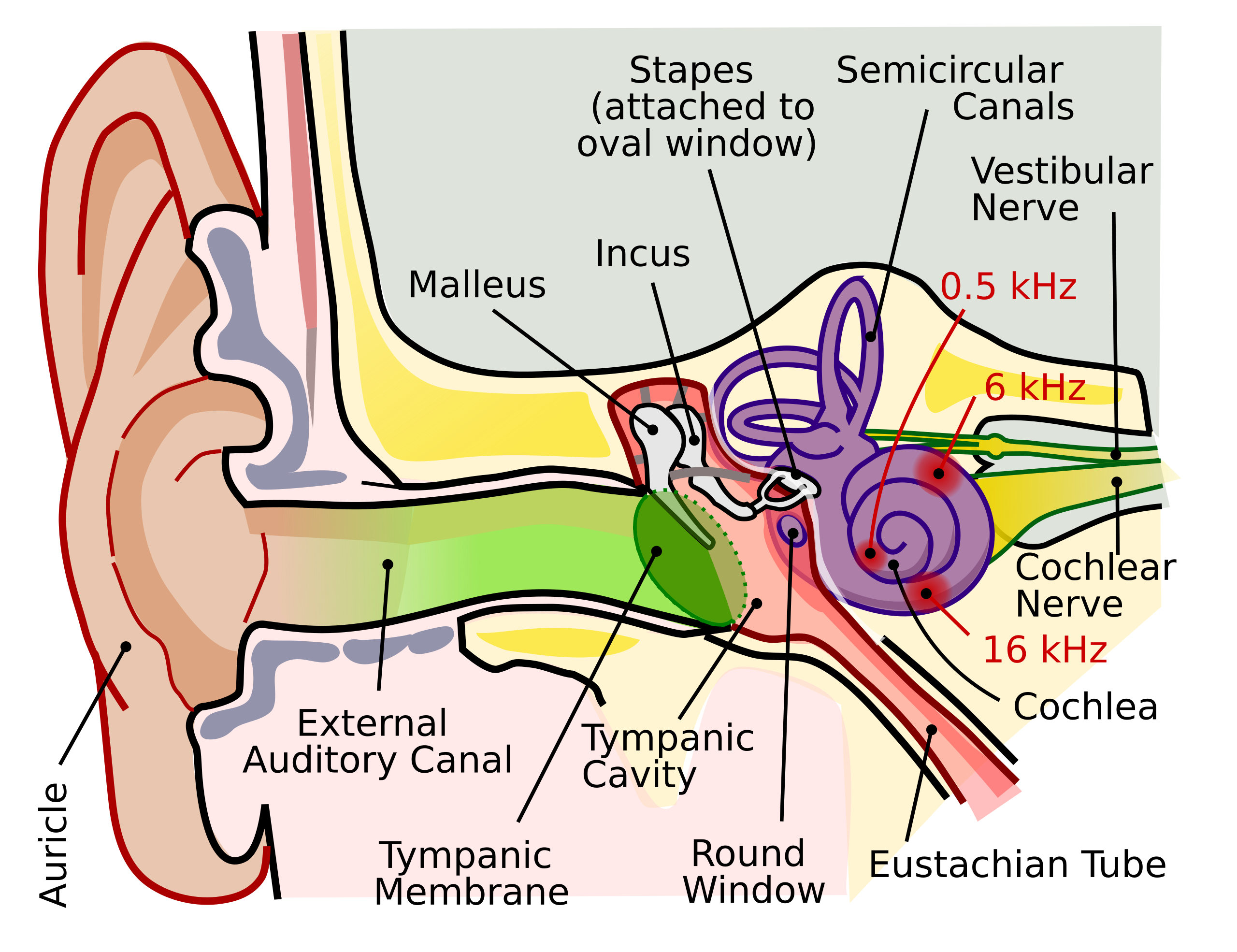
English: The human ear and frequency mapping in the cochlea. The three ossicles incus, malleus, and stapes transmit airborne vibration from the tympanic membrane to the oval window at the base of the cochlea. Because of the mechanical properties of the basilar membrane within the snail-shaped cochlea, high frequencies will produce a vibration peak near the oval window, whereas low frequencies will stimulate receptors near the apex of the cochlea (locations for three frequencies indicated schematically). Information from the cochlear receptor cells is transmitted to the cochlear nuclei via the 8th cranial nerve, and on through the midbrain to the cortex. |
| Data | |
| Origem | Obra do próprio (Texto original: “Own work by uploader, derived from File:Anatomy_of_the_Human_Ear.svg”) |
| Autor | Inductiveload |
| Permissão (Reutilizar este ficheiro) |
A utilização deste ficheiro é regulada nos termos da licença Creative Commons - Atribuição-CompartilhaIgual 2.5 Genérica.
|
| Outras versões |
[]
|
| SVG desenvolvimento | O código-fonte desta imagem SVG é válido. Este(a) desenho vetorial foi criado com o Inkscape Este arquivo é traduzido usando elementos SVG switch: todas as traduções são armazenadas no mesmo arquivo. |


Histórico do ficheiro
Clique uma data e hora para ver o ficheiro tal como ele se encontrava nessa altura.
| Data e hora | Miniatura | Dimensões | Utilizador | Comentário | |
|---|---|---|---|---|---|
| atual | 21h29min de 16 de setembro de 2018 | | 674 × 519 (33 kB) | JoKalliauer | added systemLanguage="eo" |
| 17h21min de 16 de setembro de 2018 |  | 674 × 519 (32 kB) | JoKalliauer | added systemLanguage="de" | |
| 05h33min de 11 de setembro de 2018 |  | 674 × 519 (87 kB) | Jmarchn | Bigger (proportional real size) and full redraw (more realistic) of the auricle. Ossicles in white colour. Eardrum with contour. Added 3 labels. Add fundus to the bone and subcutaneous tissues, add superior auricular muscle, add transparency to middle ear, add separation between middle and inner ear, add division to internal auditory canal. | |
| 13h40min de 29 de abril de 2009 |  | 800 × 600 (98 kB) | Inductiveload | swap incus/malleus | |
| 15h10min de 15 de fevereiro de 2009 |  | 800 × 600 (98 kB) | Inductiveload | {{Information |Description={{en|1=The human ear and frequency mapping in the cochlea. The three ossicles incus, malleus, and stapes transmit airborne vibration from the tympanic membrane to the oval window at the base of the cochlea. Because of the mechan |
Utilização local do ficheiro
Não há nenhuma página que use este ficheiro.
Utilização global do ficheiro
As seguintes wikis usam este ficheiro:
- en.wikipedia.org
- en.wikibooks.org
- eo.wikipedia.org
- he.wikipedia.org
- lt.wikipedia.org
- www.wikidata.org