Ficheiro:HIV entry into T cell schematic.png

Imagem numa resolução maior (2 805 × 3 405 píxeis, tamanho: 4,49 MB, tipo MIME: image/png)
|
|
Esta imagem provém do Wikimedia Commons, um acervo de conteúdo livre da Wikimedia Foundation que pode ser utilizado por outros projetos.
|
Descrição do ficheiro

|
Esta imagem de biologia (ou todas as imagens neste artigo ou categoria) deveriam ser recriadas usando gráficos vectoriais, como ficheiros SVG. Isto tem várias vantagens; veja as Commons:Media for cleanup|imagens para rever para mais informações. Se já criou um ficheiro SVG desta imagem, por favor, carregue-o. Depois do novo ficheiro SVG ter sido carregado, substitua aqui esta predefinição pela predefinição {{vector version available|nome da nova imagem.svg}}.
|
| Descrição |
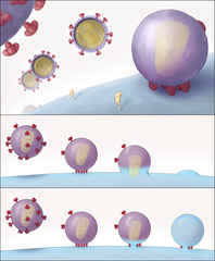
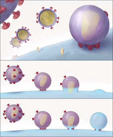
English: "Schematic Representation of the Key Structural Features of SIV and HIV-1 Entry into T Cells"
(A) Different stages of viral entry from budding, to maturation, to entry claw formation. For the SIV strain used here, viruses that are docked to the cell via an entry claw show very few, if any, viral spikes on their surface, whereas non-contacting viruses typically display between 60 and 100 spikes on their surface. The entry claw is composed of between five to seven anchors spanning the region between the virus and the cell, each ∼100 Å long, and spaced laterally by ∼150 Å. (B and C) Two alternative models for viral entry. In the global fusion model (B), the formation of the entry claw is followed by progressive fusion of the viral membrane across its width, leading to merger of the contents of the viral membrane with the cellular membrane. In the local fusion model (C), the formation of the entry claw is followed by the creation of a local pore centered at one of the rods, leading to delivery of the viral core into the cell." |
| Data | Published May 4, 2007 |
| Origem |
Sougrat R, Bartesaghi A, Lifson JD, et al (May 2007). "Electron tomography of the contact between T cells and SIV/HIV-1: implications for viral entry". PLoS Pathog. 3 (5): e63. PMID 17480119. doi:10.1371/journal.ppat.0030063 Direct link to image: http://www.plospathogens.org/article/showImageLarge.action?uri=info%3Adoi%2F10.1371%2Fjournal.ppat.0030063.g008 |
| Autor | Rachid Sougrat, Alberto Bartesaghi, Jeffrey D. Lifson, Adam E. Bennett, Julian W. Bess, Daniel J. Zabransky, Sriram Subramaniam |
| Permissão (Reutilizar este ficheiro) |
[1] |
| Outras versões | JPG version |
Licenciamento
|
A utilização deste ficheiro é regulada nos termos da licença Creative Commons - Atribuição 2.5 Genérica.
|
This file was published in a Public Library of Science journal. Their website states that the content of all PLOS journals is published under the Creative Commons Attribution 4.0 license (or its previous version depending on the publication date), unless indicated otherwise.
|
Histórico do ficheiro
Clique uma data e hora para ver o ficheiro tal como ele se encontrava nessa altura.
| Data e hora | Miniatura | Dimensões | Utilizador | Comentário | |
|---|---|---|---|---|---|
| atual | 00h30min de 11 de junho de 2008 | | 2 805 × 3 405 (4,49 MB) | Fvasconcellos | {{Information |Description="Schematic Representation of the Key Structural Features of SIV and HIV-1 Entry into T Cells" (A) Different stages of viral entry from budding, to maturation, to entry claw formation. For the SIV strain used here, viruses that |
Utilização local do ficheiro
Não há nenhuma página que use este ficheiro.
Utilização global do ficheiro
As seguintes wikis usam este ficheiro:
- ar.wikipedia.org
- en.wikipedia.org
- es.wikipedia.org
- ko.wikipedia.org
- outreach.wikimedia.org
- sl.wikipedia.org