Ficheiro:Validation of the dye diffusion assay performed with the flattened cochlear preparation.png

Dimensões desta antevisão: 287 × 599 píxeis. Outras resoluções: 115 × 240 píxeis | 230 × 480 píxeis | 979 × 2 044 píxeis.
Imagem numa resolução maior (979 × 2 044 píxeis, tamanho: 3,33 MB, tipo MIME: image/png)
|
|
Esta imagem provém do Wikimedia Commons, um acervo de conteúdo livre da Wikimedia Foundation que pode ser utilizado por outros projetos.
|
Descrição do ficheiro
| Descrição |
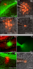
English: A–D: Dye diffusion patterns after PI was injected into a single cell in various locations in the cochlea. The type of the cells that was injected is given at lower right corner of each panel. E–F: Diffusion patterns of four different fluorescent dyes after injecting into a single Claudius cell. Name of the dye is given in the lower right corner of each panel. Panels B), C), D), F) & H) were photographed with unfixed fresh samples. Panels A), E), G) were results obtained from fixed samples after the experiments were done. They were labeled with fluorescent phalloidin (red in E, green in A&G) to outline the cell border. Scale bar on the top left of each panel represents approximately 100 µm. |
| Data | |
| Origem | PLOS ONE an open source peer reviewed journal- Gap Junction Mediated Intercellular Metabolite Transfer in the Cochlea Is Compromised in Connexin30 Null Mice ([1]) |
| Autor | Qing Chang, Wenxue Tang, Shoeb Ahmad1, Binfei Zhou1, Xi Lin1 |
Licenciamento
A utilização deste ficheiro é regulada nos termos da licença Creative Commons - Atribuição 2.5 Genérica.
- Pode:
- partilhar – copiar, distribuir e transmitir a obra
- recombinar – criar obras derivadas
- De acordo com as seguintes condições:
- atribuição – Tem de fazer a devida atribuição da autoria, fornecer uma hiperligação para a licença e indicar se foram feitas alterações. Pode fazê-lo de qualquer forma razoável, mas não de forma a sugerir que o licenciador o apoia ou subscreve o seu uso da obra.
Histórico do ficheiro
Clique uma data e hora para ver o ficheiro tal como ele se encontrava nessa altura.
| Data e hora | Miniatura | Dimensões | Utilizador | Comentário | |
|---|---|---|---|---|---|
| atual | 04h24min de 12 de fevereiro de 2009 | | 979 × 2 044 (3,33 MB) | Mike.lifeguard | {{Information |Description={{en|1=A–D: Dye diffusion patterns after PI was injected into a single cell in various locations in the cochlea. The type of the cells that was injected is given at lower right corner of each panel. E–F: Diffusion patterns o |
Utilização local do ficheiro
A seguinte página usa este ficheiro:
Utilização global do ficheiro
As seguintes wikis usam este ficheiro:
- en.wikipedia.org
- zh.wikipedia.org